We are all made up of cells. And these cells come together to form tissue, we know this. But what keeps these cells together? How does this glue form and change to promote the functions of a tissue? One approach to study this is to see how it is constructed. Investigating how this glue assembles was the focus of a collaborative project of the Mayor Lab at the National Centre for Biological Sciences, and the Lecuit Lab at the Turing Centre for Living Systems (CenTuri), France. The results from their ongoing collaboration were recently published in Current Biology.
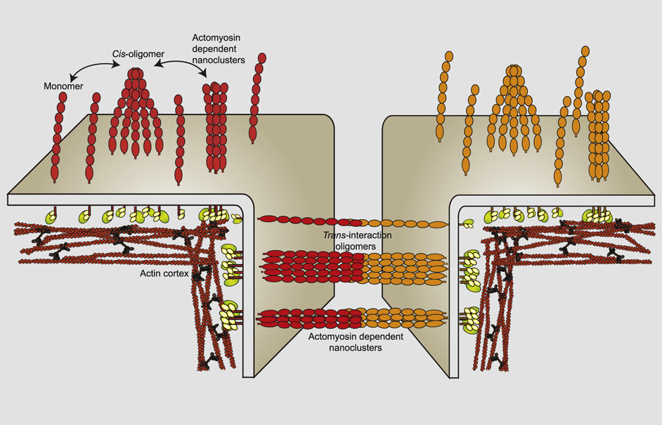
In this project, the team examined the organization and structure of Cadherins, which are a type of adhesion protein concentrated at cell-cell junctions, and necessary for the formation of tissues. In the case of epithelial tissues they are named E-cadherins, and specifically “dE-cad” refers to the E-cadherins in fruit fly (Drosophila melanogaster) epithelial cells. Cadherins are not naturally present in the Drosophila larval blood cells, so this primary cell system provided a good model system to test the role of dE-cad in cell-cell adhesion and organization. By creating their own cell-cell interaction system in the Drosophila larvae, the researchers were able to gain insights into how these adhesive proteins work. The E-cadherin system could be studied from a new perspective by this synthetic approach that is still set within the organism, using biophysical tools developed for the purpose in the Mayor Lab.
The Lecuit Lab provided and created new fly lines used in the experiments conducted at NCBS. They expressed dE-cad in the larval hemocytes, single cell systems that move around in the insect hemolymph, similarly to the immune cells in human blood. The dE-cad expression in the hemocytes was achieved through a powerful tool called the UAS-GAL4 system. They found that the organization of the cadherin protein in hemocytes mimics that of the epithelium of Drosophila embryos. With the convenience of fruit fly biology, various aspects of the cadherin system could thus be probed by utilizing specialized techniques such as FRAP (Fluorescence recovery after photobleaching), FCS (Fluorescence correlation spectroscopy) and Homo-FRET microscopy.
They found that cadherins can interact with molecules on the same cell membrane (cis interaction) as well as with molecules on the opposing membrane (trans interaction). They found that extra-junctional dE-cad (molecules present outside cell-cell junctions) are organized as immobile nanoclusters and loosely packed diffusive oligomers in hemocytes. Cis interactions of the extracellular domain of dE-cad help in generating immobile oligomers, which promotes the formation of actomyosin sensitive nanoclusters of extra-junctional dE-cad. The junctional dE-cad also exhibits trans paired nanoscale organization. “The observations suggest a hierarchical assembly mechanism for the generation of the actomyosin based nanoclusters,” says Ruma Chandran, the first author.
These findings are the product of an exciting LIA, a laboratory without walls from the CNRS, set up between Satyajit Mayor’s lab and Thomas Lectuit’s lab in 2015. Their interest in understanding biological tissue and energy use, combining principles of biology and active matter theory in physics with Madan Rao (NCBS), led them to launch a series of experiments to understand cellular organization. This study is the first of a body of work that could help elucidate how cells are dynamically controlled, and how they assemble, by understanding what happens in the constantly changing plasma membrane.
The work is built on the existing understanding of the construction and remodelling of epithelial tissue, mainly pioneered by the Lecuit lab, and a new understanding of the cell surface pioneered by the Mayor and Rao groups at NCBS. “This collaboration is an opportunity to ask how tissue dynamics emerges from the properties of cell-cell contacts, which in turn depend on active remodelling of cadherins by contractile actomyosin networks. This project stems from a sabbatical at NCBS in 2011. It became clear then that the framework developed by the Mayor and Rao labs at the nanoscale would be very useful to address junction stability and remodelling during tissue morphogenesis,” says Prof. Lecuit.
Sharing more of the history of the project, Prof. Mayor says, “Thomas and I have nurtured a shared interest in how energy consuming systems function at the molecular, cellular and tissue scales. They highlight how the glue that permits cells to interact, ‘should be considered as an out-of-equilibrium, active, and dissipative process that can be dynamically tuned by actin interactions with E-cad oligomers/nanoclusters’.This collaboration, supported by an International Research Project from the CNRS, has allowed us to sustain the work over the years. Ruma's ability to work across the two laboratories and deploy and use new tools of fly biology would not have been possible without these efforts.”
The alignment and development of these research questions has grown through an unique ecosystem where international partnerships are fostered and thrive. Dr. Srini Kaveri, Director of CNRS Bureau in India, says, “These key findings testify to the importance of international cooperation between the laboratories of two leading scientists. The collaborative International Research Project platform provided by CNRS cultivates a rewarding interaction amongst scientists and research students, and paves the path for further strengthening the bilateral relations.”


0 Comments