
Cell and tissue culture has played an important role in life science research. However, cells in culture behave quite differently from cells in vivo, and the knowledge gained from these cannot be directly extrapolated to cells in the context of an organism. There is a great interest in the scientific community to generate devices that can control the microenvironments of cells in culture.
Microfluidics devices not only allow precise control of the cellular microenvironment but also allows for economising of reagent usage. We have a state-of-the-art Microfluidics and Microfabrication Facility on our Campus that helps users to generate such devices. This facility is headed by Dr Karthik Mahesh, who also co-heads the MedTech Rapid Prototyping Facility at the Centre for Cellular and Molecular Platforms (C-CAMP).
Deepti
Hi Karthik, can you briefly tell me what your facility does?
Karthik
Our facility focuses towards fabricating structures at the micron scale. To imagine what a micron scale is, a typical hair strand is about 100 micrometres thick, and we make structures smaller than that. The size range usually goes as low as four or five microns, which is the typical size of red blood cells. In addition to fabricating microstructures, we also make microfluidic devices. Microfluidics is the phenomenon concerned with the behaviour of fluids at the micron scale. At the micron scale, the physical properties of fluids are vastly different from those at the macro scale. Simple things like Reynolds number and diffusion behave very differently at this scale. This makes it easier for biological experiments to happen in microfluidic devices.
Deepti
Interesting. My experience working with cells has been on glass coverslips and plastic dishes. What is the advantage of using these devices as compared to those methods?
Karthik
In those techniques, you use a large quantity of cells. You are culturing and experimenting with thousands of cells. Here, for the same experiment, we require only a few cells. Let's say an experiment which takes 1000 cells in a petri dish would require only about 100 cells in a microfluidic chip to get the same outcome. So, in that way, you're scaling down the quantity of reagents that you require - in terms of biological material (cells or molecules), and also the fluid/medium that you're passing through the device. It is in microlitres or, in some cases, nanolitres. That is a significant cost reduction. And also, at that scale, like I said, the properties of the fluids behave differently. Let's take ELISA, for example. If it takes 30 minutes for an incubation step in conventional ELISA, it takes only a few minutes in a microfluidic device. You're saving on time, quantities, materials, and cost.
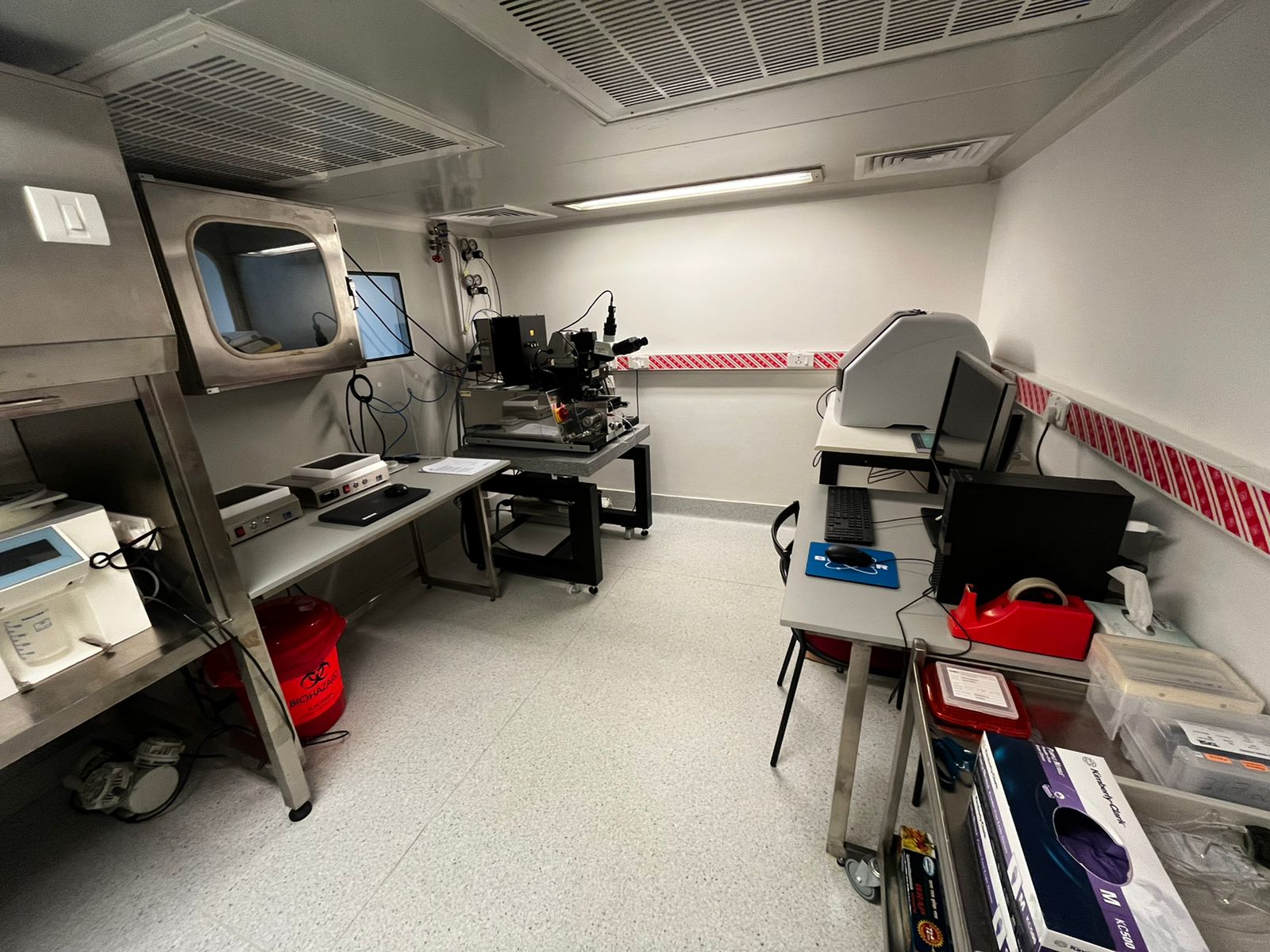
The tools required for fabricating micron-scale features are housed in a Class 10000 cleanroom that has low contamination levels.
Deepti
So, what kind of material is used to make these devices? Are they similar in some way to biological substrates, or are they closer to glass and plastic?
Karthik
The substrate that we use is actually a silicon wafer. Microfabrication technology is derived from the microelectronics industry. Our cell phones and laptops have integrated circuits which are essentially made of silicon wafers. On top of the silicon wafer, we coat something called a photoresist, which is a compound that is sensitive to light. Before that, we create “stencils”, or a template that has the micro-scale features that you want. We place this template on top of the photoresist and allow UV light to pass through it. The regions that are exposed to light get hardened up, and the remaining regions get washed away. What you're left with on your silicon substrate is your microstructure. On top of that, we pour a material called PDMS (Polydimethylsiloxane). PDMS is a material that you will find on the ingredient list of many shampoos and hairsprays. It's actually used for creating lustre in these products. But here, we use PDMS for making these microfluidic devices. So, once we pour and cure the PDMS, it becomes very soft and elastic. It's also a very biocompatible material, which is why we use it for biological experiments. Also, it's optically transparent with a refractive index close to that of glass. Once PDMS is cured, we cut the device out from the silicon wafer and bond it to a glass substrate using a device called a plasma cleaner. We can then attach some tubing to flow biological samples through the chip; the device is ready at this stage.
Deepti
Cells adhere and spread on coverslips and may not behave the same way in the body. Is it different in these devices?
Karthik
We're essentially trying to mimic a biological system using microfluidic devices. So, we have to design the device in such a way that it can mimic the microscale structure of a biological system. For example, the skin has different layers composed of various cell types. Using microfluidic structures, we can build those layers in such a way that once it is seeded with cells and the culture medium is introduced through it, the complete device will mimic actual organs or tissues.
Deepti
That is very interesting. So could you tell me some of the achievements of the facility or devices that you have made for specific experiments?
Karthik
We regularly work with the Discovery to Innovation Accelerator (DIA) at C-CAMP, for whom we supply a flow cytometer device. Typically, flow cytometers are large, and they take up a lot of space. This particular flow cytometer can essentially fit in the palm of your hand.
Apart from that, we get regular requests from clients, mostly academic institutions. So recently, we made a microarray device for a client from VIT, Vellore. They wanted a microarray with 6-micron features for studying the properties of red blood cells. We were able to successfully fabricate and deliver such devices.
In addition to these, we have been training a lot of individuals through workshops that we conduct. Microfluidics technology is still a very nascent technology yet to catch up amongst the scientific community. So, what we're trying to do is propagate this technology to as many people as we can through such events.
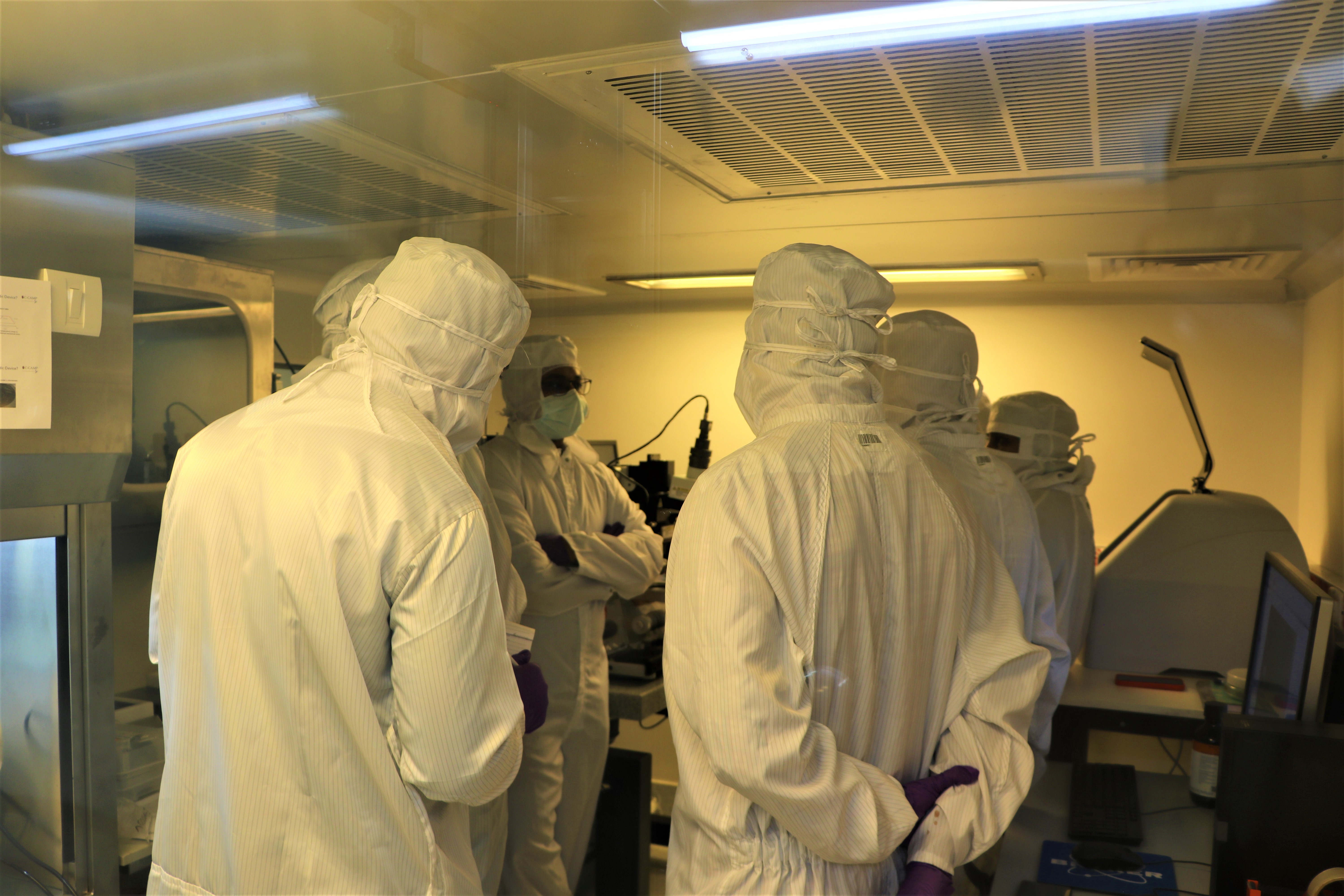
Karthik (centre) can be seen here training users on various aspects of microfabrication and microfluidics in the facility's cleanroom.
Deepti
Very nice. Karthik, what led you to pursue this field?
Karthik
I am a Bangalorean and did my Bachelor's and Master's in Biotechnology from M S Ramaiah Institute of Technology. For a college project, I was working with animals (mostly rats and mice) and doing some drug screening. I felt so sorry for these animals. I then came across an article which said that microfluidics technology through organs-on-chips can replace animal testing. So, that's where this technology sort of fascinated me. I remember that from that point onwards, all my classroom presentations during my Master’s were all focused on something related to microfluidics. My teachers realized that I was completely obsessed with this field. I then decided to do a PhD. My PhD was from the Centre for Nanoscience and Engineering (CeNSE) at the Indian Institute of Science (IISc), and I graduated in 2021. My thesis was focused on microfluidic impedance cytometry, which is an electrical way of characterizing individual cells in flow. We were able to characterize the electrical and mechanical properties of blood cells using a microfluidic device integrated with microelectrodes.
Deepti
That's very fascinating. What are some of the special skills that you bring to your current job from your training?
Karthik
Coming from a Biotechnology background and being in a department like CeNSE has been very useful for my current work profile. At CeNSE, the research is mostly focused towards electronics applications and semiconductor fabrication. As I mentioned before, microfabrication technology is derived from the electronics industry, so the tools that you use for semiconductor processing are the same tools used for microfluidics too. The kind of exposure that I got working in a cleanroom environment has been extremely beneficial. Working in a cleanroom with tools used for lithography, diffusion, thin film deposition, wet etching, and dry etching has given me a special skill set in microfabrication. Because my thesis was focused on microfluidics, I was able to gain a good foundation on the fundamental aspects of fluid flow at the micro-scale. In summary, I bring an interdisciplinary skill set ranging from fundamental biology to microfabrication to microfluidics.
Deepti
So Karthik, what are your hobbies? And when not working, what do you like to do?
Karthik
I got married recently, and my wife is a surgeon at one of the leading medical colleges in Bangalore. So, when I’m not working, I try to spend as much time with my wife as I can since she gets very limited time from her job. We try to explore places in and around Bangalore as much as we can. Apart from this, I enjoy traveling and reading books.
Deepti
What is your favorite genre in books?
Karthik
Mostly fiction, fantasy and sci-fi.

Karthik enjoys traveling. This picture is from a recent trip to Mysore.
Deepti
Yeah. So, as you said already, there is limited knowledge of these devices in the scientific community. So how do you see the future of this technology?
Karthik
I think microfluidics is a vast subject with various applications. One application that is already being used right now is for diagnostics. For example, when you go to a diagnostic centre for a blood test, they require at least 5 mL of blood. Microfluidics technology will enable you to do the same test with just a single drop of blood. This will have a tremendous impact on the cost of diagnostic techniques and the manpower involved. And of course, the goal is to replace the conventional gold standards of biochemical tests for diagnosis.
Another major use that I already mentioned is for replacing animal testing. Recently, many American companies have already declared a ban on animal testing for cosmetics. We are slowly moving towards replacing animal testing with organ-on-a-chip devices made using microfluidics and microfabrication techniques. Apart from that, usage in fundamental biological research is going to increase. In experiments that involve miniaturization, such technologies can help provide new insights into biological phenomena that we are still unaware of.
Deepti
So, final question. If somebody wants to explore or use these devices, how should they approach you?
Karthik
They can contact me via e-mail and discuss their specific requirements. External users can approach the facility through C-CAMP. We provide various services, including designing, fabricating, and testing these devices. We have specialized software for designing microfluidic devices and microstructures which individuals may utilize. We also offer technical consultations on any aspect of microfluidics and microfabrication; be it design, fabrication or experimentation.


0 Comments