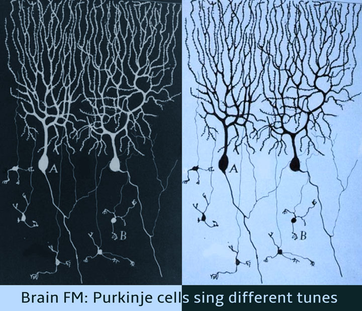
Brain FM: Purkinje cells sing different tunes
Each of us have our moods when we like to hum an old melody or suddenly feel like tapping our feet to the latest hit. It turns out that cells in our brains can be equally moody, changing the tune of their electrical signals from time to time. In a recent study, scientists Mohini Sengupta and Dr. Vatsala Thirumalai, from the National Centre for Biological Sciences (NCBS), Bangalore, reveal that nerve cells found in the cerebellum (at the base of the brain) send out electrical signals in either a constant hum or in sudden bursts. Which of the two tunes they choose depends on the voltage across their cell membranes and on input from a specific region of the brain.
Our balance, co-ordination and the capacity to learn new skills such as riding a bicycle or playing a piano depends on a small leaf-like structure at the base of our brain called the cerebellum. Within the cerebellum, are nerve cells named 'Purkinje cells' that are arranged neatly in a single layer and are vital for carrying out the aforementioned functions. These cells receive signals from many different regions of the brain and send out messages to the deeper layers of the cerebellum.
However, how Purkinje cells communicate with other nerve cells has thus far been a mystery, mainly because it is very difficult to 'listen' to these cells in animals that are awake and moving around. Since these nerve cells are very small, scientists employ fine glass capillaries to record their electrical signals using a technique called 'whole cell patch clamping'. For this technique to work, most experimental animals must be anesthetised, as even small movements can knock the capillaries out of place. Unfortunately, anesthetics themselves alter the electrical signals generated by the brain. This means that previous studies aimed at determining the nature of electrical signals generated by Purkinje cells in awake and moving animals have largely been inconclusive.
The researchers at NCBS overcame these issues by using zebrafish, a fresh water fish found in the Ganga and Brahmaputra, for their experiments. The young zebrafish (called larvae) are transparent and have not yet developed a skull. Furthermore, specific nerve cells in the larvae can be made to glow by injecting DNA into them at the embryonic stage. This made it possible for scientists to insert and precisely place the fine recording equipment onto the Purkinje cells within the fish brains. To prevent the fish from moving, a paralytic agent that does not interfere with electrical signals in the brain was used. This combination of technical advances allowed the team to record electrical signals from Purkinje cells in an intact animal that was not anesthetized.
Results from these experiments showed that Purkinje cells sent out electrical signals in two different modes depending on the voltage at their cell surfaces. The first mode, called the 'down' state, occurred when the inside of the cell was more negative compared to the outside. In this state, cells were silent until signals from a different part of the brain arrived, at which time, they sent out a burst of impulses. In the second mode, called the 'up' state, the inside of the cell was less negative compared to the outside and the Purkinje cells sent out impulses at a constant rate. In this mode, these cells ignored any impulses coming from other parts of the brain. These states are reminiscent of a person attentively listening to directions and responding accordingly, or simply 'zoning out' and going their own way irrespective of instructions.
What does this phenomenon mean for the functioning of the cerebellum? To understand this, the NCBS scientists recorded the instructions of the nervous system to muscles. In normal fish, these instructions would result in swimming. However, since the experimental preparation is paralyzed, no movement is seen, yet the instructions to the muscle continue to be sent. The scientists discovered that such 'fictive' motor instructions were always accompanied by bursts of impulses in the Purkinje cells. Mostly, the impulses occurred after the start of these motor instructions and the timing of the bursts varied from cell to cell.
These results demonstrate that Purkinje cells receive a copy of the instructions sent by the nervous system to muscles and that they generate a burst of impulses in response. The researchers propose that the existence of the 'up' and 'down' states is a possible mechanism by which Purkinje cells choose to listen in on the instructions sent out to muscles or not. What do these cells do with such instruction copies? Do they alter their signals when the animal is learning a new motor task? The authors are excited to find out answers to these questions in ongoing experiments.
The paper titled "AMPA receptor mediated synaptic excitation drives state-dependent bursting in Purkinje neurons of zebrafish larvae" was published in the journal eLife on 29th September 2015 and can be accessed here.

Comments
Post new comment