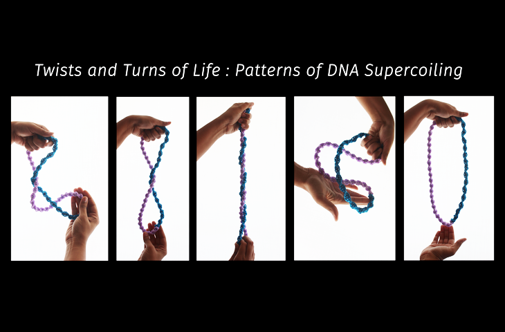
"A bend and a twist, then stretch and turn, now relax". What sounds like a series of exercise instructions, are also words that describe the various shapes a piece of DNA can assume. The classic double helix structure that one associates with DNA is but an extremely limited view of its physical 'shape'. The molecule that holds the codes of life is capable of further winding itself into myriad complex shapes called 'supercoils' that are capable of affecting gene expression patterns. Now, researchers from the National Centre for Biological Sciences (NCBS), Bangalore, and the National Institutes of Health (NIH), USA, have elucidated this pattern of supercoiling across the genome of the much studied bacterium E. coli.
DNA molecules are wound and rewound into complex structures that condense their immense lengths to a fraction of their actual size in order to fit their long strings of information into microscopic cells. But this 'packed' DNA that fits neatly into a cell also needs to be 'unpacked' periodically for gene expression and duplication. Transcription and replication require localised sections of DNA to be unwound from its double helix. These activities cause further twisting and coiling or 'overwinding' in regions of DNA elsewhere. Therefore, a battery of DNA-binding proteins such as NAPs (Nucleoid-Associated Proteins), transcription factors or those involved in replication, and enzymes like toposiomerases keep a cell's genetic material in a constant state of structural and functional equilibrium. Coils, supercoils, bends, twists and turns are formed, lost and reformed depending on a cell's state of activity.
A bacterial cell can be exposed to various environmental changes which include periods of starvation, lack of oxygen and unfavourable temperatures. Surviving these situations would require the bacterium to alter genetic expression profiles. Scientists have long thought that these changes could be effected through variations in the supercoiled structure of DNA. For example, the genomes of actively dividing cells under rich-nutrient conditions are known to be more negatively supercoiled than the genomes of cells from the stationary phase when nutrients are scarce. In other words, supercoiling is likely to be sensitive to changes in the environment.
Although recent advancements in methodology have allowed researchers to study DNA supercoiling in eukaryotic cells at local scales, this methodology has never been applied to prokaryotes. Researchers from Aswin S. N. Seshasayee's group at NCBS and Prof. Sankar Adhya's team at NIH have recently applied these methods to study DNA supercoiling in bacteria at a fine-scale level. Using the chemical trimethylpsoralen, exposure to UV light and microarray technology, the research team have gained information on section-specific variations in genomic supercoiling within bacteria exposed to different external conditions.
"We have measured DNA supercoiling at a fine-scale resolution in bacteria for the first time. This study provides proof-of-concept that the supercoiling of a genome is not uniform and that it varies locally across genes. It also provides evidence to support the hypothesis that bacterial cells could be regulating gene expression and their own physiologies by altering the structure of their genomes," says Avantika Lal, the first author of the publication in the journal Nature Communications that details these findings.
In order to study the effect of environmental stimuli on the supercoiling status of the bacterial genome, two populations of E.coli were used to simulate two different external conditions. One simulated a nutrient-rich situation where actively dividing cells represented a growing population; whereas the other represented a condition where a population had exhausted its nutrients and was in a 'stationary' phase. Since the binding of trimethylpsoralen to DNA is proportional to the amount of supercoiling in the DNA, one could study genome-wide patterns of winding under these two settings.
The results have shown that E. coli cells in the stationary phase display a gradient of supercoiling across their circular genomes, with the lowest supercoiling at a region named 'ori' and the highest supercoiling at a region named 'ter'. The 'ori' (short for 'origin') region refers to the area in the genome where many genes essential for bacterial reproduction are encoded. The 'ori' region is also where DNA replication begins or originates, whereas 'ter' (short for 'terminus') refers to the area in the genome where replication terminates. Furthermore, the gradient of supercoiling in stationary phase cells was found to be dependent on the protein 'Hu' which is a well-known NAP. In actively dividing cells, however, such a gradient was missing. This result essentially refutes the hypothesis that negative supercoiling in dividing cells would be higher at the 'ori' than the 'ter' to favour origin-proximal gene transcription and initiation of replication.
"This was a surprise result. We believe that the higher activity of DNA gyrase and higher levels of transcription cause the genome-wide increase in negative supercoiling in actively dividing cells," says Avantika. "We hope to understand these processes in future studies that include inhibiting DNA gyrase and transcription activity in these situations. It is very early days yet, but this work paves the way to understanding which genes' expression are affected by the environment, and how we could control cell physiology through them by altering external conditions," she adds.
This work has been published in the paper titled "Genome scale patterns of supercoiling in a bacterial chromosome" in March 2016 in the journal Nature Communicatioons. The paper can be accessed here.


0 Comments